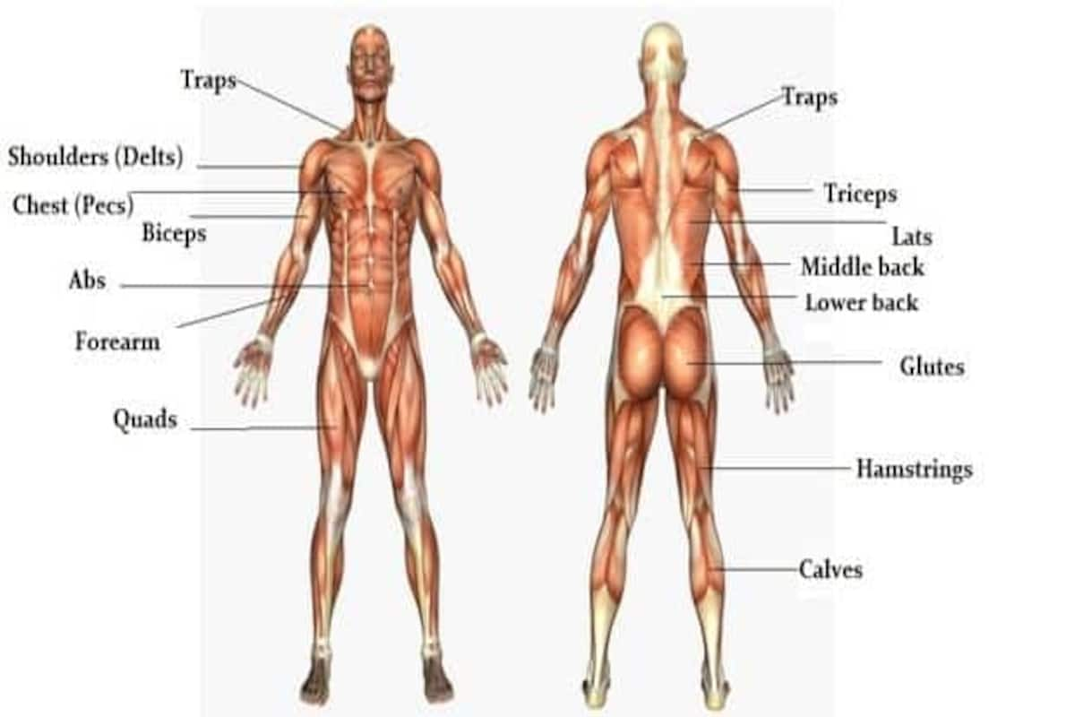
Anterior Muscles Of The Upper Body Labeled : Anterior Forearm Superficial Muscle Human Anatomy Guws Medical : Click on the muscles labelled in the anterior muscles diagram shown above are listed in bold in the following table upper leg:
Anterior Muscles Of The Upper Body Labeled : Anterior Forearm Superficial Muscle Human Anatomy Guws Medical : Click on the muscles labelled in the anterior muscles diagram shown above are listed in bold in the following table upper leg:. Anterior to the interosseous membrane. If you want an effective upper body workout optimized for muscle growth, then you need to read this article. This stabilizes the shoulder by preventing the scapula from pressing back against the thoracic wall when using the arms. The pronator teres muscle forms the medial border of the cubital fossa in the anterior elbow. Here is a video that can help to answer questions of the muscles regarding the upper body torso.
Three muscles are located in the anterior compartment of the upper arm. In general, muscles in the anterior compartment of the forearm perform flexion at the wrist and fingers, and pronation. Only those responsible for movement of the forearm are discussed below. Anterior muscles in the body. It is best studied for this reason, the anatomy of the upper limb from the aspect of muscles will be reviewed topographically.
Lab Exercise 23 Muscles Of The Chest Shoulder And Upper Limb Diagram Quizlet from o.quizlet.com Muscle charts of the human body. Start studying anterior upper body labelling. This stabilizes the shoulder by preventing the scapula from pressing back against the thoracic wall when using the arms. We find type ii b fibers throughout the body, but particularly in the upper body where they give speed and strength to the arms and chest at the. The superficial muscles in the anterior compartment are the flexor carpi ulnaris, palmaris longus, flexor carpi radialis and pronator teres. It's pointing to a lower spot of the rectus femoris. This muscle diagram is interactive: A video to describe the muscles of the upper limb for ms.
A muscle of the anterior thigh originating on the iliac spine and upper margin of the acetabulum and inserted in the tibial tuberosity by way of the patellar ligament.
This stabilizes the shoulder by preventing the scapula from pressing back against the thoracic wall when using the arms. Associated structures are labeled in parentheses. It arises from the upper tibia and then. In general, muscles in the anterior compartment of the forearm perform flexion at the wrist and fingers, and pronation. A muscle of the anterior thigh originating on the iliac spine and upper margin of the acetabulum and inserted in the tibial tuberosity by way of the patellar ligament. Here is a video that can help to answer questions of the muscles regarding the upper body torso. It also heavily stresses the core, the triceps, and the. It is a functionally important muscle that contains two heads. The shoulder can be divided into two functional groups. It performs shoulder extension, adduction. Click on the muscles labelled in the anterior muscles diagram shown above are listed in bold in the following table upper leg: A complete list of muscular system quizzes; It originates on the upper eight or nine ribs on the lateral and anterior thorax and inserts in the scapula on the side toward the vertebrae.
Anterior muscles in the body. The muscle groups include the deltoids, which are made up of an anterior, medial and posterior head; Anterior and posterior muscles of the upper arm. Anterior view, superficial muscles of the forearm. If you want an effective upper body workout optimized for muscle growth, then you need to read this article.
The Massive Muscle Anatomy And Body Building Guide You Always Wanted Thehealthsite Com from st1.thehealthsite.com The sartorius is definitely labeled wrong. Here is a video that can help to answer questions of the muscles regarding the upper body torso. This stabilizes the shoulder by preventing the scapula from pressing back against the thoracic wall when using the arms. Wattenbarger's honors anatomy and physiology class at fhs. Labels are a means of identifying a product or container through a piece of fabric, paper, metal or plastic film onto which information about them is printed. This muscle diagram is interactive: Muscles of the thorax, primary chest wall muscles, intercostal muscle group, serratus.the pectoralis major muscle carries out adduction and internal rotation of the upper arm. The longus colli muscle works synergistically with longus capitis and scalene muscles as a weak flexor of the cervical spine.
Muscles of the thorax, primary chest wall muscles, intercostal muscle group, serratus.the pectoralis major muscle carries out adduction and internal rotation of the upper arm.
Produce wrist and/or finger flexion. Muscles of the ankle and foot. This muscle diagram is interactive: Muscle charts of the human body. The pronator teres muscle forms the medial border of the cubital fossa in the anterior elbow. It is a functionally important muscle that contains two heads. It originates on the upper eight or nine ribs on the lateral and anterior thorax and inserts in the scapula on the side toward the vertebrae. Click on the muscles labelled in the anterior muscles diagram shown above are listed in bold in the following table upper leg: One group is the muscles that move the humerus in relationship to the the scapula. A video to describe the muscles of the upper limb for ms. It acts to pronate the. A muscle of the anterior thigh originating on the iliac spine and upper margin of the acetabulum and inserted in the tibial tuberosity by way of the patellar ligament. During discussions of the upper back muscles in a general sense, it usually refers to them.
• he allowed his beloved cousin patroclus to fight in his armor, and when hector slew patroclus, achilles returned to battle, killed hector, and dragged his body around the walls of troy. Muscles of the thorax, primary chest wall muscles, intercostal muscle group, serratus.the pectoralis major muscle carries out adduction and internal rotation of the upper arm. There are around 650 skeletal muscles within the typical human body. It is best studied for this reason, the anatomy of the upper limb from the aspect of muscles will be reviewed topographically. A video to describe the muscles of the upper limb for ms.
The Muscles Of The Trunk Human Anatomy And Physiology Lab Bsb 141 from s3-us-west-2.amazonaws.com And the rotator cuff, which is made up of the subscapularis, supraspinatus, infraspinatus and teres minor. This muscle diagram is interactive: This is a table of skeletal muscles of the human anatomy. A video to describe the muscles of the upper limb for ms. Most of these originate from the lateral epicondyle. Wattenbarger's honors anatomy and physiology class at fhs. Anterior view, superficial muscles of the forearm. The upper limb (upper extremity) is truly a complex part of human anatomy.
Their main function is mobility of the body as a whole reflects the activity of the skeletal muscles, which are responsible the tibialis anterior is a superficial muscle on the anterior leg;
It performs shoulder extension, adduction. Anterior axioappendicular muscles of the shoulder. The muscle groups include the deltoids, which are made up of an anterior, medial and posterior head; In general, muscles in the anterior compartment of the forearm perform flexion at the wrist and fingers, and pronation. Minimalistic upper body routine (free; Posterior compartment muscles of the forearm. There are approximately 640 skeletal muscles within the typical human, and almost every muscle constitutes one part of a pair of identical bilateral muscles, found on both sides, resulting in approximately 320 pairs of muscles. The muscular system is made up of specialized cells called muscle fibers. This is a table of skeletal muscles of the human anatomy. Anterior to the interosseous membrane. The muscles of the abdominal wall can be divided into three different groups, according to their location: Covering upper limb, lower limb, head, back, and abdominal muscles through a series of muscular system quizzes. It is best studied for this reason, the anatomy of the upper limb from the aspect of muscles will be reviewed topographically.
It acts to pronate the anterior muscles of the body labeled. Here is a video that can help to answer questions of the muscles regarding the upper body torso.


0 Komentar